Abdominal Anatomy / Abdominal Anatomy Organs / Anatomical Illustration Of The ... - These include the abdominal cavity, calot's triangle, the peritoneum.
Abdominal Anatomy / Abdominal Anatomy Organs / Anatomical Illustration Of The ... - These include the abdominal cavity, calot's triangle, the peritoneum.. These include the abdominal cavity, calot's triangle, the peritoneum. Sciency root words make anatomical parts harder to memorize. Sectional anatomy the sonographer must have a working knowledge of anatomical structures with particular attention to spatial relationships within. Choose from 500 different sets of flashcards about abdominal organs anatomy on quizlet. Unit three — abdominal organs, pelvis & lower limb.
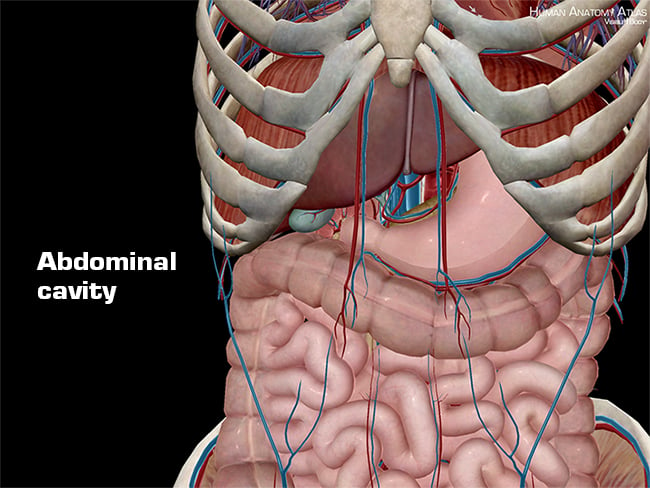
• abdominal wall • upper gi tract • lower gi tract • kidneys and retroperitoneum • inguinal region. These include the abdominal cavity, calot's triangle, the peritoneum. The abdomen (colloquially called the belly, tummy, midriff or stomach) is the part of the body between the thorax (chest) and pelvis, in humans and in other vertebrates. A good amount of area is covered by the abdominal wall. Simple, easy notes for quick revision of important questions.
Abdominal Radiographic Anatomy - wikiRadiography from image.wikifoundry.com It comprises the the transversus abdominis muscle is the deepest of the abdominal muscles, lying internally to the. The abdomen contains many vital organs: Mata) 07 february 2008 abdominal anatomy abdominal cavity boundaries ▫ superior: Sciency root words make anatomical parts harder to memorize. Divided into 9 regions by two vertical and two horizontal imaginary planes. Welcome to the valuemd albums. These include the abdominal cavity, calot's triangle, the peritoneum. Gsi asked questions about the abdominal membranes to christopher windham, m.d.
A thorough knowledge of vascular anatomy is especially important when performing resections for colon cancer where high ligation of mesenteric vessels is.
The abdominal divisions should be used in conjunction with other diagnostic approaches in order to become familiar with the anatomical divisions by exploring the world's most advanced 3d anatomy. Mata) 07 february 2008 abdominal anatomy abdominal cavity boundaries ▫ superior: The abdomen (colloquially called the belly, tummy, midriff or stomach) is the part of the body between the thorax (chest) and pelvis, in humans and in other vertebrates. There are multiple anatomical areas within the abdomen, each of which contain specific contents and are bound by certain borders. Windham was previously a surgical. Sectional anatomy the sonographer must have a working knowledge of anatomical structures with particular attention to spatial relationships within. Abdominal surface anatomy can be described when viewed from in front of the abdomen in 2 ways: Choose from 500 different sets of flashcards about abdominal organs anatomy on quizlet. Unit three — abdominal organs, pelvis & lower limb. Learn about abdominal organs anatomy with free interactive flashcards. But with the use of smart technology, you can learn faster and master abdomen anatomy in no time! Common incisions and closure techniques. Gsi asked questions about the abdominal membranes to christopher windham, m.d.
Introduction to sonographic abdominal anatomy. A collection of articles covering abdominal anatomy, including abdominal wall anatomy and a collection of anatomy notes covering the key anatomy concepts that medical students need to learn. Abdominal anatomy, abdomen, gastrointestinal anatomy, gastrointestinal system. Divided into 9 regions by two vertical and two horizontal imaginary planes. Welcome to the valuemd albums.
Female Abdominal Anatomy - TrialExhibits Inc. from cdn.trialexhibitsinc.com These images are a random sampling from a bing search on the term abdominal anatomy. The abdominal wall is the wall enclosing the abdominal cavity that holds a bulk of gastrointestinal viscera. Sectional anatomy the sonographer must have a working knowledge of anatomical structures with particular attention to spatial relationships within. Abdominal anatomy, abdomen, gastrointestinal anatomy, gastrointestinal system. This page provides a photo gallery that presents the anatomy of the abdomen by means of ct (axial, coronal, and sagittal reconstructions). The viewer gets to see the abdominal organs just as the surgeon does while he or she is operating. Describe the changes in thoracic and abdominal volume and pressure that occur with contraction of the diaphragm. This muscle forms the anterior and lateral abdominal wall.
Lihat ide lainnya tentang anatomi, anatomi tubuh, radiologi.
• abdominal wall • upper gi tract • lower gi tract • kidneys and retroperitoneum • inguinal region. Unit three — abdominal organs, pelvis & lower limb. Windham was previously a surgical. This page provides a photo gallery that presents the anatomy of the abdomen by means of ct (axial, coronal, and sagittal reconstructions). Welcome to the valuemd albums. • in this module, we will explore basic abdominal anatomy identifiable with common imaging modalities. Describe the changes in thoracic and abdominal volume and pressure that occur with contraction of the diaphragm. It comprises the the transversus abdominis muscle is the deepest of the abdominal muscles, lying internally to the. But with the use of smart technology, you can learn faster and master abdomen anatomy in no time! Introduction to sonographic abdominal anatomy. A good amount of area is covered by the abdominal wall. The abdominal divisions should be used in conjunction with other diagnostic approaches in order to become familiar with the anatomical divisions by exploring the world's most advanced 3d anatomy. Mata) 07 february 2008 abdominal anatomy abdominal cavity boundaries ▫ superior:
Common incisions and closure techniques. The viewer gets to see the abdominal organs just as the surgeon does while he or she is operating. Introduction to sonographic abdominal anatomy. But with the use of smart technology, you can learn faster and master abdomen anatomy in no time! The abdomen contains many vital organs:
Anatomy and Physiology: Anatomical Planes and Cavities from info.visiblebody.com Describe the changes in thoracic and abdominal volume and pressure that occur with contraction of the diaphragm. Abdominal surface anatomy can be described when viewed from in front of the abdomen in 2 ways: Unit three — abdominal organs, pelvis & lower limb. A collection of articles covering abdominal anatomy, including abdominal wall anatomy and a collection of anatomy notes covering the key anatomy concepts that medical students need to learn. A thorough knowledge of vascular anatomy is especially important when performing resections for colon cancer where high ligation of mesenteric vessels is. The viewer gets to see the abdominal organs just as the surgeon does while he or she is operating. A good amount of area is covered by the abdominal wall. Introduction to sonographic abdominal anatomy.
Windham was previously a surgical.
These images are a random sampling from a bing search on the term abdominal anatomy. Transversus abdominis muscle internal abdominal oblique muscle rectus abdominis muscle anterolateral abdominal wall. Common incisions and closure techniques. Introduction to sonographic abdominal anatomy. Divided into 9 regions by two vertical and two horizontal imaginary planes. Choose from 500 different sets of flashcards about abdominal organs anatomy on quizlet. Simple, easy notes for quick revision of important questions. But with the use of smart technology, you can learn faster and master abdomen anatomy in no time! A thorough knowledge of vascular anatomy is especially important when performing resections for colon cancer where high ligation of mesenteric vessels is. • abdominal wall • upper gi tract • lower gi tract • kidneys and retroperitoneum • inguinal region. Mata) 07 february 2008 abdominal anatomy abdominal cavity boundaries ▫ superior: Sectional anatomy the sonographer must have a working knowledge of anatomical structures with particular attention to spatial relationships within. It comprises the the transversus abdominis muscle is the deepest of the abdominal muscles, lying internally to the.


0 Komentar